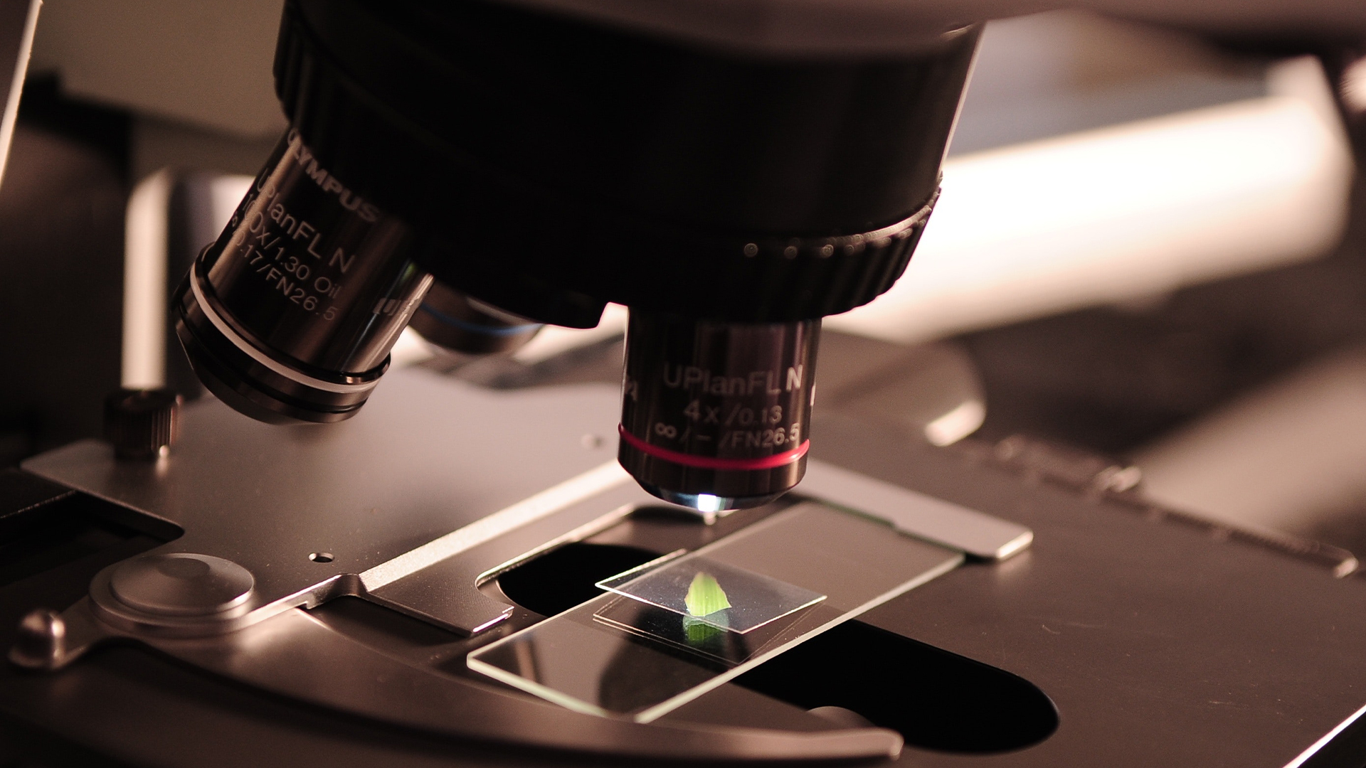
What connects the Covid-19 vaccine, police forensics departments and the latest generation of computer processing chips? None of them would have been possible without microscopes.
Microscopy, a cornerstone of scientific discovery, is being transformed by AI and automation, driving significant advancements across multiple fields. Industries from pharmaceuticals to software rely on microscopy to support operations and drive innovation.
Despite its critical role, the microscopy workflow – from image acquisition to analysis and generating actionable insights – has traditionally been complex and time-consuming.
Today’s life scientists are harnessing automation and artificial intelligence (AI) technologies to radically streamline the process. Let’s take a closer look at how this technology will affect each step of the process.
Streamlining image acquisition
Acquiring a workable, high-quality image of the item under inspection requires manually calibrating numerous software and hardware parameters. This is both time- and labour-intensive and demands specialised domain knowledge. AI-driven automation now streamlines this process by automatically adjusting these parameters, reducing the need for constant manual input.
Furthermore, sometimes parameters need to be changed while image acquisition is underway. For instance, if the object under examination is growing or altering its position under the lens, the settings will have to shift with it. This requires vigilance and agility from a human operator, creating potential for error and bias. AI can dynamically respond, adjusting settings in real-time
Another advantage of digitised microscopy with AI integration is the image metadata it creates. The final file will contain information on the image, plus the microscope and camera settings that produced it. This provides valuable material for the analysis stage. It also makes experiments much easier to reproduce.
Enhancing image analysis
Once the microscope has produced a set of quality images, the process of detailed analysis and interpretation begins.Automation can then prepare the image for close analysis. Image enhancement software will remove noise and sharpen the definition for a crisp and clear picture. But the real AI magic happens at the segmentation stage.
Much medical research and clinical activity depends on detecting and classifying the contents of microscope images. For instance, when diagnosing cancer, researchers will need to be able to distinguish between normal and cancerous cells in a sample.
Done manually, this requires researchers to painstakingly inspect and annotate multiple datasets. However, the application of AI transforms this process. Researchers need only manually annotate a single dataset with the correct segments and then present it to a deep learning model for training. The model, now trained on the taxonomy, can automatically differentiate between segments in subsequent samples. This performs in seconds what could take researchers hours freeing them up for higher value creation and increasing a lab’s productivity.
Automating output creation
The final stage of the microscopy pipeline is to turn analysis into outputs. Scientific reports require a great deal of precise information and so manual creation can be arduous – which is where automation can help.
A report is normally accompanied by a log of all activities performed during the microscopy process, information about errors and technical details. Using automation, these factors can be recorded during acquisition and analysis stages, and compiled into a comprehensive metadata log.
With high-quality images also automatically stored on disk, ready for insertion throughout the report, many ingredients of a professional research output no longer require human labour, but instead rely on human oversight to ensure accuracy and reliability in results.
Human oversight for AI-powered scientific research
AI could revolutionise microscopy’s capabilities. But not every stage in every experiment should be automated. Human input and oversight are integral to the process and extremely valuable. The endless potential to customise digital microscopes gives researchers the freedom to find the optimal structure for every project.
We’ve been pleased to support ZEISS Microscopy as it works to empower researchers with AI-powered tools. For the full story, read our case study.
Or, for more ways AI is changing the healthcare industry, download our whitepaper: Unlocking the Potential of Generative AI in Healthcare and Life Sciences.
This article has been updated by Endava since it was originally published.
